Lab Report by Yi Lee
Name: Choong Yi Lee 111359Lab 1 Principles and Use of Microscope
1.1 Setting up and using the microscope
Introduction: In order to be seen,
microorganisms need to be magnified. Despite advances in other area of
microscopy (for example, the electron microscope), the light microscope is
still the instrument most frequently used for viewing microorganisms.
Objective: Learn to use a simple bright-field microscope correctly.
Results:
Total magnification = objective lens power x eyepiece lens
power(10x)
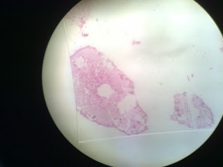
Typical bacillus
40x magnification
40x magnification
100x magnification
400x magnification
Discussion
on Typical Bacillus
1 The sample specimen is observed under the
magnification of 40x, 100x and 400x
2 The light intensity is adjusted to ensure
that the sample specimen can be observed clearly.
3 The stage, fine adjustment knob and coarse
adjustment knob are also adjusted to get a clear observation of specimen.
Morphology
a) Color- red
b ) Surface- smooth in appearance
c) Shape- irregular in appearance
d) Texture- dry
e) Elevation- flat
Conclusion: The specimen can be seen clearly and
obviously under the magnification on 400x. In this experiment, we can conclude
that the higher the power of magnification, the clearer the observation and the
better result.
Reference: -System
Microbiology, Edited by: Brian D. Robertson and
Brendan W. Wren
-Bacteria Spores, Edited by: Ernesto Abel-Santos
-http://faculty.ccbcmd.edu/courses/bio141/lecguide/unit1/shape/shape.html
-http://www.microbiologybytes.com/video/Bacillus.html
-http://www.microbiologybytes.com/video/Bacillus.html
1.2 Examination of cells
Introduction:
Because of their extreme minuteness, bacteria are not generally studied
with the lower-power or high power-power dry objectives. Instead they are
stained and observed with the oil immersion objective.
The wet mount methods enables you
to study the sizes and shapes of living microorganisms (drying or staining
microorganism distort them). It also enables you to determine if cells are
motile. The wet mount method is quick and easy, and does not require special
equipment.
Objective:
- To provide an experience in the use of microscope.
- To provide an experience in the use of microscope.
-To illustrate the diversity of cells and microorganisms.
Results:
Discussion
1 The oil immersion oil is put on the the top of specimen
2 The power magnification of 1000x is adjusted to view and sticking to the specimen
3 Adjust the fine adjustment knob so that can get a clearer view of the specimen.
2 The power magnification of 1000x is adjusted to view and sticking to the specimen
3 Adjust the fine adjustment knob so that can get a clearer view of the specimen.
Morphology
Lactobacillus:
a)Shape-rods with rounded end
b) Color-yellowish
c) Texture- Moist
b) Color-yellowish
c) Texture- Moist
d) Size- Small and Tiny
a) Form in shape- circular/globular
b) Elevation- flat
b) Elevation- flat
c) Texture- moist
d) Surface- dull
e) color- violet
f) size- small and tiny rod shape
f) size- small and tiny rod shape
Reference: -http://en.wikipedia.org/wiki/Saccharomyces
-http://www.biology.ed.ac.uk/archive/jdeacon/microbes/yeast.htm
- http://www.ehow.com/facts_5792238_physical-description-lactobacillus- acidophilus.html
- http://www.ehow.com/facts_5792238_physical-description-lactobacillus- acidophilus.html


No comments:
Post a Comment